Toxo in Canadian deer hunters eating undercooked venison from Illinois

We conducted a recent investigation in Quebec, Canada, concerning Canadian deer hunters who went to the United States to hunt deer and returned with symptoms of fever, severe headache, myalgia, and articular pain of undetermined etiology. Further investigation identified that a group of 10 hunters from Quebec attended a hunting retreat in Illinois (USA) during November 22–December 4, 2018.

Six of the 10 hunters had similar symptoms and illness onset dates. Serologic tests indicated a recent toxoplasmosis infection for all symptomatic hunters, and the risk factor identified was consumption of undercooked deer meat. Among asymptomatic hunters, 2 were already immune to toxoplasmosis, 1 was not immune, and the immune status of 1 remains unknown. Outbreaks of acute toxoplasmosis infection are rare in North America, but physicians should be aware that such outbreaks could become more common.

Acute toxoplasmosis among Canadian deer hunters associated with consumption of undercooked deer meat hunted in the U.S.

Emerging Infectious Diseases, vol. 26, no. 2

Colette Gaulin , Danielle Ramsay, Karine Thivierge, Joanne Tataryn, Ariane Courville, Catherine Martin, Patricia Cunningham, Joane Désilets, Diane Morin, and Réjean Dion

https://wwwnc.cdc.gov/eid/article/26/2/19-1218_article?deliveryName=DM17555

TB in deer hunters

For a country that still proclaims, we enjoy the safest food supply in the world in U.S. Department of Agriculture missives, when we’ve been arguing reduced risk is a better message for 25 years and that there are so many countries with the self-proclaimed title of safest food in the world they can’t all be right – it’s alarming that Mycobacterium bovis has been transmitted from deer to a human.

Hello zoonoses.

Deer hunting season in Ontario (that’s in Canada) begins about today.

I never had any interest.

Not a Bambi thing, just thought it was boring.

My dad went a few times but I’m not sure if he enjoyed it or not.

Whatever.

The U.S. Centers for Disease Control reports that in May 2017, the Michigan Department of Health and Human Services was notified of a case of pulmonary tuberculosis caused by Mycobacterium bovis in a man aged 77 years. The patient had rheumatoid arthritis and was taking 5 mg prednisone daily; he had no history of travel to countries with endemic tuberculosis, no known exposure to persons with tuberculosis, and no history of consumption of unpasteurized milk. He resided in the northeastern Lower Peninsula of Michigan, which has a low incidence of human tuberculosis but does have an enzootic focus of M. bovis in free-ranging deer (Odocoileus virginianus). The area includes a four-county region where the majority of M. bovis–positive deer in Michigan have been found.

Statewide surveillance for M. bovis via hunter-harvested deer head submission has been ongoing since 1995; in 2017, 1.4% of deer tested from this four-county region were culture-positive for M. bovis, compared with 0.05% of deer tested elsewhere in Michigan. The patient had regularly hunted and field-dressed deer in the area during the past 20 years. Two earlier hunting-related human infections with M. bovis were reported in Michigan in 2002 and 2004. In each case, the patients had signs and symptoms of active disease and required medical treatment.

Whole-genome sequencing of the patient’s respiratory isolate was performed at the National Veterinary Services Laboratories in Ames, Iowa. The isolate was compared against an extensive M. bovis library, including approximately 900 wildlife and cattle isolates obtained since 1993 and human isolates from the state health department. This 2017 isolate had accumulated one single nucleotide polymorphism compared with a 2007 deer isolate, suggesting that the patient was exposed to a circulating strain of M. bovis at some point through his hunting activities and had reactivation of infection as pulmonary disease in 2017.

Whole-genome sequencing also was performed on archived specimens from two hunting-related human M. bovis infections diagnosed in 2002 (pulmonary) and 2004 (cutaneous) that were epidemiologically and genotypically linked to deer (3). The 2002 human isolate had accumulated one single nucleotide polymorphism since sharing an ancestral genotype isolated from several deer in Alpena County, Michigan, as early as 1997; the 2004 human isolate shared an identical genotype with a grossly lesioned deer harvested by the patient in Alcona County, Michigan, confirming that his infection resulted from a finger injury sustained during field-dressing. The 2002 and 2017 cases of pulmonary disease might have occurred following those patients’ inhalation of aerosols during removal of diseased viscera while field-dressing deer carcasses.

In Michigan, deer serve as maintenance and reservoir hosts for M. bovis, and transmission to other species has been documented. Since 1998, 73 infected cattle herds have been identified in Michigan, resulting in increased testing and restricted movement of cattle outside the four-county zone. Transmission to humans also occurs, as demonstrated by the three cases described in this report; however, the risk for transmission is understudied.

Similar to Mycobacterium tuberculosis, exposure to M. bovis can lead to latent or active infection, with risk for eventual reactivation of latent disease, especially in immunocompromised hosts. To prevent exposure to M. bovis and other diseases, hunters are encouraged to use personal protective equipment while field-dressing deer. In addition, hunters in Michigan who submit deer heads that test positive for M. bovis might be at higher risk for infection, and targeted screening for tuberculosis could be performed. Close collaboration between human and animal health sectors is essential for containing this zoonotic infection.

Notes from the Field: Zoonotic mycobacterium bovis disease in deer hunters—Michigan, 2002-2017

James Sunstrum, MD1; Adenike S hoyinka, MD2; Laura E. Power, MD2,3; Daniel Maxwell, DO4; Mary Grace Stobierski, DVM5; Kim Signs, DVM5; Jennifer L. Sidge, DVM, PhD5; Daniel J. O’Brien, DVM, PhD6; Suelee Robbe-Austerman, DVM, PhD7; Peter Davidson, PhD5

https://www.cdc.gov/mmwr/volumes/68/wr/mm6837a3.htm

‘He would be dead right now’ Canadian family warns parents after 2-year-old contracts E. coli infection

There is so much I want to write about, to get the daily buzz of the blog, but blogs don’t last, and don’t pay the bills.

What’s important to me is the pic, below, and everything I can do to help them succeed.

I’ll work on books (which also don’t pay the bills, but may last longer).

Julian Kolsut of Chek reports that a Parksville family is at the B.C. Children’s Hospital in Vancouver this weekend after their two-year-old toddler contracted an E. coli Infection — that his father Aaron Hughes says was not diagnosed until it was almost too late.

“Be persistent… if we didn’t stick to our guns and react, he would be dead right now,” said Hughes.

It all started when 2-year-old Jaxon starting to act “off” on Tuesday, both of his parents thought he may have had heatstroke and made sure to take him out of the sun and keep him hydrated.

“He was out in the sun and he became really lethargic really fast, didn’t have an appetite, a fever kicked in quickly and then the diarrhea started,” said Aarons wife Jolene Secord.

After contacting the nurse’s line, they were given the same advice, but eventually Jaxon started vomiting. A visit to the walk-in clinic resulted in a different diagnosis, a possible bacterial infection, but while at the clinic they noticed blood in his diaper.

“They did take a swab of his green bowl movements but sent us home,” said Secord.

They were told to continue keeping an eye on him and taking care of him.

The parents say on Wednesday the vomiting and bleeding got worse, and after rushing Jaxon to the Nanaimo Regional General Hospital they were told again it may be a bacterial infection, and that he may have had a torn fissure. After speaking to a nutritionist they were sent home.

Aaron says he couldn’t see the fissure, and was confused the mucus coming out of his toddler’s behind was also bloody.

When his 18-year-old daughter called him when he was shopping for vaseline for his son in Nanaimo on Thursday and was told Jackson’s symptoms were worse, he told them to call and ambulance at that he would meet them at the hospital.

“We weren’t wasting any time… but they couldn’t find his stools [that were sent in for testing]” said Hughes.

“The doctor then came into the room [the next day] and said, “I think we found the source” and he didn’t look too happy, [the doctor] tracked down the stools and said the test came back positive for E. coli O157:H7.”

He was immediately airlifted to the B.C. Children’s Hospital in Vancouver and currently, among many other issues with his health, does not have any functioning kidneys and has a damaged pancreas.

Jaxon is undergoing dialysis, transfusion and other treatments, and his mom says his state changes by the hour.

“At this point, they don’t see the kidneys waking up… it could be a long-term thing… every hour seems to change for Jaxon,” said Secord. “We will see a positive, we will see a negative, we will see a positive.”

The exact cause is unknown, but the family says they suspect it came from deer feces, as the animal can carry the O157:H7 strain. They think Jackson may have come in contact with it outside.  Contaminated food is also a possibility.

“We almost have a family of deer living [on our property]” said Hughes. “We have apple trees on our property and they [the deers] live off that stuff, and that can transport diseases.”

Both parents also say the unfortunate incident is a reminder to always listen to your gut.

“If we would have stuck to what they were telling us then we would have lost him… and if we waited he could have had brain damage, heart failure,” said Hughes.

DCIM102GOPROGOPR2675.JPG

Canadian E. coli cases believed to have been caused by deer meat

Cured deer meat is believed to be behind a series of E. coli cases in Tavistock, Oxford County, in Ontario, Canada.

Public Health says they can’t confirm it yet, but they believe the illnesses were caused by the meat which was sourced and processed from two private hunt camps in December 2017.

The first case was reported by a Tavistock resident in mid-February with the second coming a month later in March and a third in the first week of April.

They say laboratory results, expected later this week, will confirm if E.coli is present in the deer meat.

‘Disease from outer space’ States confront the spread of CWD in deer

In March, 1996, the UK government confirmed what had been known for years: bovine spongiform encephalopathy (or mad cow disease) was killing humans in the UK.

The various forms of transmissible encephalopathies have different names according to the species – scrapie in sheep, feline spongiform encephalopathy in cats, Creutzfeldt-Jacob disease in humans, chronic wasting disease in deer and elk.

But they’re all the same affliction, caused by infectious proteins, or prions.

I haven’t been following the CWD outbreak in deer, but it seems to be where BSE was about 1993: There’s this mysterious new disease no one ever thoughts would cross over to humans, but now, maybe?

Jim Robbins of the New York Times writes that, as darkness closed in, one hunter after another stopped at this newly opened game check station, deer carcasses loaded in the beds of their pickups.

They had been given licenses for a special hunt, and others would follow. Jessica Goosmann, a wildlife technician with Montana’s Fish, Wildlife & Parks Department, stepped outside to greet them, reaching for the neck of each freshly killed deer to cut an incision and remove a lymph node for testing.

On the edge of this south-central Montana village, where deer hunting is a way of life, the game check station has become the front line of the state’s efforts to stop the spread of a deadly infection known as chronic wasting disease.

It has ravaged deer herds throughout the United States and Canada and forced the killing of thousands of infected animals in 24 states and three Canadian provinces. It has also been found in Norway and South Korea. With the disease widespread in Wyoming, the Dakotas and the province of Alberta, Montana officials had been bracing for its emergence.

So in November, when biologists discovered it in six deer in this part of Montana and in another near the Canadian border, officials began setting up special hunts and stations for testing.

“It wasn’t a surprise that we found it,” said John Vore, game management bureau chief for the Montana Department of Fish, Wildlife & Parks. “It was a disappointment, but not a surprise.”

On Friday, the department announced that two more deer from this region, taken early in the special hunt, tested positive for the disease. Other test results are pending.

Chronic wasting disease is a contagious neurological disease that infects elk, deer, moose and caribou, and reduces their brains to a spongy consistency. Animals become emaciated, behave strangely and eventually die. It’s not known to be transferred to humans. Neither is it known to be spread from wild to domestic animals. There is no treatment, although a vaccine has been successful in tests in wild deer.

It is among a class of diseases known as transmissible spongiform encephalopathy, or TSE. Most experts believe the infectious agent is something called a prion, a misfolded cellular protein found in the nervous system and lymph tissue. The disease was first noted in captive deer in Colorado in the 1960s. The most closely related animal disease is scrapie in sheep.

“It’s a very unusual disease,” said Matthew Dunfee, an expert at the Wildlife Management Institute in Fort Collins, Co. and project director for the Chronic Wasting Disease Alliance. “Some experts say it’s a disease from outer space.”


10 men ‘violently’ ill after wild venison meal

Deer meat, or venison, was a staple at Amy’s dinner table.

Me, never liked it, preferred grain-fed beef.

Guess I’m just a suburban kid.

A Brookfield, Wisconsin,  man says he and 10 of his friends all became violently ill after sharing a meal of home-cooked wild venison, but what made them sick remains a mystery.

Scott Mathison said the symptoms didn’t appear for five days after his church retreat to Black River Falls October 1st.

“I could feel the life leaving my body. I knew something was something really serious,” Mathison said Friday. “I was violently shaking, had a 104 (degree) fever when my wife took me in to urgent care. If I wouldn’t have been treated, I’m not sure I would’ve came through that.”

While the symptoms included night sweats and high fevers, the lack of gastro-intestinal issues convinced Mathison that he didn’t have food poisoning. And he later learned that he wasn’t alone in feeling ill.

“I found out one of the other guys was sick, and then I found another one was sick and so we started calling and checking and we were all having the exact same symptoms, and we realized we didn’t have the flu,” he said.

Mathison said their doctors still don’t know what ailed the men, but antibiotics seem to help.

Bambi poops in water, 4 kids get sick with E. coli O157, 2016

In May 2016, an outbreak of Shiga toxin–producing Escherichia coli O157 infections occurred among children who had played in a stream flowing through a park. Analysis of E. coli isolates from the patients, stream water, and deer and coyote scat showed that feces from deer were the most likely source of contamination.

In the United States, recreational water is a relatively uncommon source of Shiga toxin–producing Escherichia coli (STEC) O157 outbreaks (1). We describe an outbreak of STEC O157 infections among children exposed to a contaminated stream in northern California, USA, and provide laboratory evidence establishing wildlife as the source of water contamination.

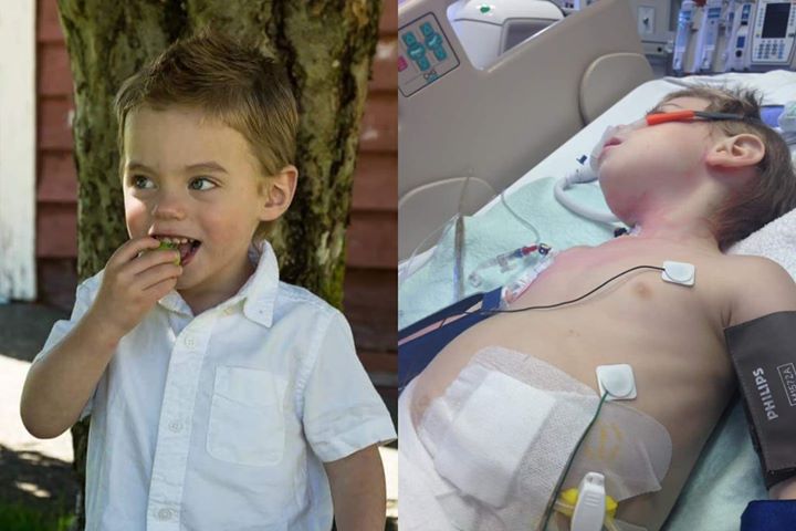
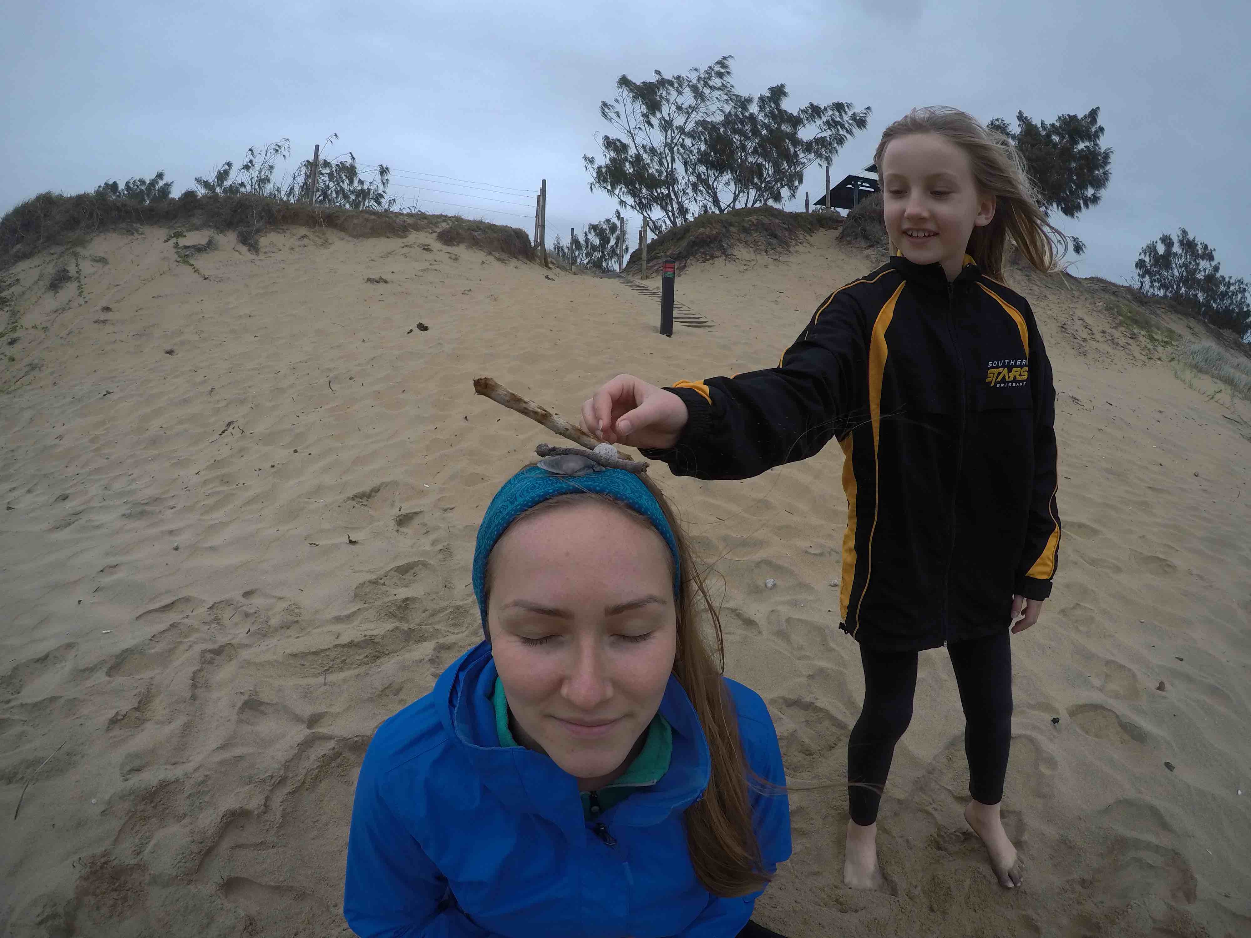
In May 2016, four cases of Shiga toxin (Stx) 1– and 2–producing E. coli O157 infection were reported to a local health department in northern California; investigation revealed a common source of exposure. The case-patients, ranging in age from 1 to 3 years, had played in a stream adjacent to a children’s playground within a city park. Exposure of the case-patients to the stream occurred on 3 separate days spanning a 2-week period. Two case-patients are known to have ingested water while playing in the stream. Two case-patients were siblings. All case-patients had diarrhea and abdominal cramps; bloody diarrhea was reported for 3. One case-patient was hospitalized with hemolytic uremic syndrome.

The stream is a second-order waterway located in a northern California community of ≈7,500 residents. At the time of exposures, stream flow was <30 ft3/s. The land upstream is not used for agricultural activities such as livestock production. The community is serviced by a public sewer system; inspection of sewer lines indicated no breach to the system.

Water samples were collected from the exposure site 7 days after the last case-patient was exposed and weekly thereafter for 17 weeks; samples were tested quantitatively for fecal indicator organisms. Throughout the study period, all water samples exceeded recreational water quality limits for E. coli and enterococci levels (2). Water samples were also cultured for STEC isolation and PCR detection of stx1 and stx2 (3). Stx1- and Stx2-producing E. coli O157 were isolated from stream water each week for the first 4 weeks. Additionally, an Stx2-producing E. coli non-O157 strain was isolated from the stream in the first week of sampling. Enrichment broth cultures of water samples were also positive by PCR for stx1 and stx2 for the first 4 weeks of sampling. Thereafter, both stx1 and stx2, or stx2 only, were intermittently detected in enrichment broth cultures for 9 additional weeks.

In the absence of an obvious source (e.g., upstream agricultural operation or sewer leak), wildlife was considered as a possible contributor to water contamination. Thirteen fresh wildlife scat specimens were collected along the stream for STEC culture and PCR. Of the 13 scat specimens, 8 originated from deer, 2 from raccoon, and 1 each from coyote, turkey, and river otter. Six scat specimens (4 deer, 1 coyote, 1 river otter) were positive for stx1 and stx2 or for stx2 by PCR (Technical Appendix[PDF – 16 KB – 1 page]). Stx1- and Stx2-producing E. coli O157 were isolated from deer scat and coyote scat. An Stx2-producing E. coli non-O157 strain was isolated from a deer scat specimen. The animal origin of the coyote and river otter scat specimens were definitively identified by partial DNA sequencing of mitochondrial cytochrome b (4).

To assess strain relatedness, we compared STEC O157 isolates from the case-patients, water, deer scat, and coyote scat by using pulsed-field gel electrophoresis (PFGE) and multilocus variable-number tandem-repeat analysis (MLVA) (5). PFGE patterns for XbaI-digested genomic DNA were highly similar among all isolates; only slight variations were found in the lower-sized bands (Figure). PFGE patterns for genomic DNA samples digested with BlnI also demonstrated a high degree of similarity (data not shown). Furthermore, MLVA profiles were identical for the case-patient, water, and deer scat isolates; only the coyote scat isolate differed from the main profile by 2 repeats at a single locus (VNTR_3).

This study provides laboratory evidence linking STEC O157 infections with the ingestion of recreational water that was probably contaminated by wildlife scat. Wild ruminants, including deer and elk, are known carriers of STEC and have been connected to outbreaks of human infections (69). We detected STEC in 50% of deer scat specimens collected from the stream bank. One of these specimens, found 1.5 miles upstream of the exposure site, contained an E. coli O157 isolate that was highly similar by molecular subtyping to case-patient and water isolates. These findings support the likelihood that feces from deer carrying STEC were the source of water contamination or, at the very least, contributed to the persistence of STEC in the water. It is unknown whether the STEC detected in coyote and river otter scat represents carriage or transitory colonization within these animals.

The common risk factor among the case-patients in this STEC O157 outbreak was exposure to a natural stream within a city park. After the outbreak was recognized, signs warning of bacterial contamination were posted along the stream. No further STEC O157 infections attributed to stream water exposure were reported.

Dr. Probert is the assistant director for the Napa-Solano-Yolo-Marin County Public Health Laboratory. His research interests focus on the development of molecular diagnostic tools for the detection of infectious agents.

Acknowledgment

We thank Frank Reyes, Keith Snipes, and Nailah Souder for their technical assistance; the County of Marin Health and Human Services and Environmental Health Services for information about the epidemiologic and environmental investigation; and the Microbial Diseases Laboratory Branch of the California Department of Public Health and the Santa Clara County Public Health Laboratory for the molecular subtyping data.

References

Heiman KE, Mody RK, Johnson SD, Griffin PM, Gould LH. Escherichia coli O157 outbreaks in the United States, 2003–2012. Emerg Infect Dis. 2015;21:1293–301. DOIPubMed

United States Environmental Protection Agency. 2012. Recreational water quality criteria. Office of Water 820-F-12–058 [cited 2017 Apr 13]. https://www.epa.gov/sites/production/files/2015-10/documents/rwqc2012.pdf

Probert WS, McQuaid C, Schrader K. Isolation and identification of an Enterobacter cloacae strain producing a novel subtype of Shiga toxin type 1. J Clin Microbiol. 2014;52:2346–51. DOIPubMed

Parson W, Pegoraro K, Niederstätter H, Föger M, Steinlechner M. Species identification by means of the cytochrome b gene. Int J Legal Med. 2000;114:23–8. DOIPubMed

Hyytia-Trees E, Lafon P, Vauterin P, Ribot EM. Multilaboratory validation study of standardized multiple-locus variable-number tandem repeat analysis protocol for Shiga toxin–producing Escherichia coli O157: a novel approach to normalize fragment size data between capillary electrophoresis platforms. Foodborne Pathog Dis. 2010;7:129–36. DOIPubMed

Fischer JR, Zhao T, Doyle MP, Goldberg MR, Brown CA, Sewell CT, et al. Experimental and field studies of Escherichia coli O157:H7 in white-tailed deer. Appl Environ Microbiol. 2001;67:1218–24. DOIPubMed

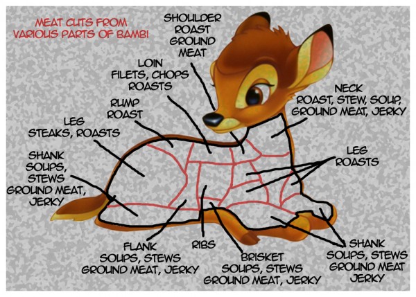
Keene WE, Sazie E, Kok J, Rice DH, Hancock DD, Balan VK, et al. An outbreak of Escherichia coli O157:H7 infections traced to jerky made from deer meat. JAMA. 1997;277:1229–31. DOIPubMed

Rounds JM, Rigdon CE, Muhl LJ, Forstner M, Danzeisen GT, Koziol BS, et al. Non-O157 Shiga toxin–producing Escherichia coli associated with venison. Emerg Infect Dis. 2012;18:279–82. DOIPubMed

Laidler MR, Tourdjman M, Buser GL, Hostetler T, Repp KK, Leman R, et al. Escherichia coli O157:H7 infections associated with consumption of locally grown strawberries contaminated by deer. Clin Infect Dis. 2013;57:1129–34. DOIPubMed

 Contaminated stream water as source for Escherichia coli O157 illness in children

05.may.17

William S. Probert, Glen M. Miller, and Katya E. Ledin

Emerging Infectious Diseases, vol. 23, no. 7, July 2017

https://wwwnc.cdc.gov/eid/article/23/7/17-0226_article
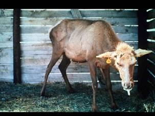
NZ dad isn’t fazed at all by the outrage at his daughter’s first deer kill

John Edens of Stuff reports a proud father says he isn’t worried by critics upset at photographs of his eight-year-old daughter taking a bite out of a deer’s heart after her first successful hunt.

kid.deer.heart.2Johnny Yuile and his daughter went hunting on a friend’s bush block the Hawke’s Bay, earlier this month.

Yuile – a New Zealand police constable – said he has been hunting with his daughter since she was a toddler and when she was little he’d strap her to a front-pack during bush missions.

His daughter, using her dad’s Remington 7mm-08 rifle, shot a young stag from 40 metres.

He sent a couple of photographs online to a Facebook page, showing a smiling father and daughter beside the stag and his daughter taking a bite from the deer’s heart. For many people, tasting a freshly killed animal, whether it’s by taking a bite or drinking blood, is part of hunting.

Yuile said he hunts for meat for his family.

“She made tricky downhill shot using my shoulder as a leaning rest and shot with dads 7 mm-08 at about 40m. Then she took a bite from its warm quivering heart,” a Facebook post said.

The Facebook page quickly attracted “haters” who criticised Yuile for taking his daughter hunting at such a young age, branded the hunt “sadistic” and wrongly accused the pair of killing an animal for sport.

Yuile said the pair were hunting at a friend’s place.

Half are children: 40 hospitalized after anthrax outbreak in Russia

The Siberian Times reports that Russian army biological protection troops have been called in amid warnings of ‘utmost care’ needed to stop anthrax from spreading.

deer.anthrax.russia.jul.16The concern among experts is that global warming thawed a diseased animal carcass at least 75 years old, buried in the melting permafrost, so unleashing the disease. 

A total of 40 people, the majority of them children, from nomadic herder families in northern Siberia are under observation in hospital amid fears they may have contracted the anthrax. Doctors stress that so far there are no confirmed cases. 

Up to 1,200 reindeer were killed either by anthrax or a heatwave in the Arctic district where the infection spread.

Specialists from the Chemical, Radioactive and Biological Protection Corps were rushed to regional capital Salekhard on a military Il-76 aircraft. 

They were deployed by Defence Minister Sergei Shoigu to carry laboratory tests on the ground, detect and eliminate the focal point of the infection, and to dispose safely of dead animals.

The 40 are all from a total of 63 nomads belonging to a dozen families who were at the site of the outbreak at Tarko-Sale Faktoria camp. The remaining nomads have been evacuated some 60 kilometres from the focus of infection in Yamalsky district.

A prolonged period of exceptionally hot weather in an Arctic Siberian district – with temperatures of up to 35C – has led to melting of permafrost in Yamalo-Nenets and other regions. 

The outbreak of anthrax earlier this week is the first in this part of Russia since 1941.

Officials say 1,200 reindeer have died in recent days, evidently through a combination of infection from anthrax, and the heatwave – unprecedented in living memory.

A major inoculation programme is also underway with a local state of emergency declared at Tarko-Sale Faktoria camp, above the Arctic Circle and close to the Yaro To lake, some 340 kilometres north-east of Salekhard.

Cats eating dead deer behind UK market stall

Officers investigating concerns about the state of a market stall found cats eating a dead deer when they went to inspect the premises.

market.stallHusband and wife, Charles, 78 and Margery Todd, 77, who sold food at markets across the East Riding as well as Leeds, Malton and York, have been banned from running any food business by magistrates, who described the images as “harrowing”.

The Todds, who ran Rosedale Farm Products, Bewholme, Hornsea, will also have to pay almost £15,000 after admitting a string of food safety and hygiene offences.

Beverley Magistrates were told the business had been operating for 48 years and supplied gamebirds, chicken and turkey to the private and commercial sector as well to the public via market stalls.

There were no hand washing facilities or provision for cleaning or washing of preparation tables or equipment.

As the meat had not been stored correctly, some was so decomposed officers were unable to identify what animal it was.

Magistrates were told how the Todds agreed to a voluntary closure order but continued to operate the business.

Mr Todd apologised to customers today saying there was “no defence” from the pictures and saying standards had slipped following two motor accidents which had affected his mobility and led to his being registered disabled.

He said: “I am not going to be bloodyminded. It was a mess. It was a gradual deterioration proportionate to the deterioration of my physical ability.”